-
Home

-
Human Medicine
-
Surgery
- Artikel


")
Surgery |
|||
Patient transport, 1921 |
|||
As early as 1830, the Bern hospitals had a travel cart for transporting island patients to the Aargau baths. Starting in 1870, Christian MIESEN made the production of large-scale tour wagons in Bonn, soon followed by the first paramedics for horse-riding. In 1899, the "Frankfurter Freiwillige RettungsGesellschaft" replaced their mobile carriers with horse-drawn carriages. Other cities were hesitant: a city like Bielefeld did not make its first horse-drawn ambulance until 1907.
In 1903, for the first time, the city of Luxembourg seems to have sought a public regulation of the transport of the sick and injured and to have obtained cost estimates. In the city council was at the beginning of 1903 (Saturday 17.1.1903) discussed on the subject. In the summer of 1903 a loan was granted:
From a statement of costs from the year 1914 it may be concluded that this ambulance was already equipped with rubber tires before the First World War:
A picture taken around 1921 is presented. From right to left:
From 1918, the Luxembourg state put a car ambulance in service. The permission to use the car had to be obtained in each case at the Ministry. In a "Circulaire" of 29.6.1918 the Minister asked for restraint at the request of the car "eu égard à la rareté et a la cherté des pneus" (quoted: A. Praum, Edm. Knaff, Code médical 1919 p. 251 ). The city of Luxembourg followed the example of 1928. In 1937, the municipality of Escher finally managed a motorized ambulance. |

Surgery |
||
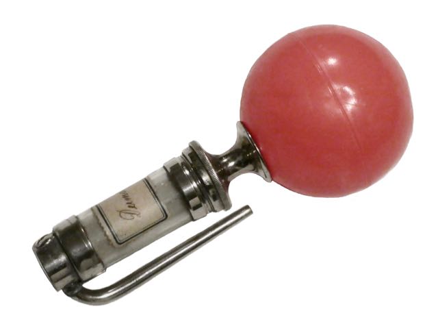
Powder distributor by GERSUNY |
||
Robert Gersuny (1844-1924) was an Austrian surgeon. He is considered the inventor of the paraffin injections, with which he created "subcutaneous prostheses" that replaced lost tissue components. To do this, he injected a multi-ointment, Vaseline, under the skin and observed that if left untouched for a time, the injected mass remained in place without irritation, without being significantly absorbed by the body , The fact that there were few traces left on the outer skin led him to apply the method to a whole range of different forms of disease, most of which involved masking the loss of tissue constituents. However, this was a momentous mistake, because over the next few decades, it has been found that the injection of paraffins can in many cases lead to lipogranulomas.
"Powdery means are most conveniently blown into the ear with a powder blower." Among the instruments which can be used for this purpose, the powder blower shown in Fig. 30, which was modified according to the powder blower of Gersuny specified by Mosetig and Wölfler, seems to me to be most handy For the purpose of ear treatment, I had the powder blower set up so that it can be handled with pipes projecting at right angles from the powder room "(Victor Urbantschitsch, Lehrbuch der Ohrenheilkunde, Verlag Urban & Schwarzenberg, Berlin, Vienna 1901).
exhibit Powder blower n. GERSUNY with bayonet-type cannula, from the collection of a general practitioner in Halle (flea market harbor in Innsbruck, 9/2018). On the glass cylinder sticks a label with the handwritten text "Dermatol". The rubber balloon was missing when purchased and was reshaped for recording by means of a small plastic ball.
Pictures in the - Catalog of the "Austro-German rubber factory defeat Gustav Berger, Vienna IX, Kolingasse 4 (1909, p.2, Fig.8). - Waldek & Wagner. Price book on surgical and medical instruments and aids, bandages, orthopedic machines and artificial limbs, dressings, apparatus for the care and relief of the sick. (1905 p.93, Fig.1483).
To dermatol "A new wound healing agent intended to replace iodoform has recently been discovered by two researchers from Wroclaw, Dr. Liebrecht and Heinz, dermatol, a body containing bismuth, which, like iodoform, is in the form of a fine yellow powder But whereas iodoform, as we know, has an unbearable, penetrating odor, which is noticeable even at great distances, Dermatol is completely odorless and, in contrast to its predecessor, it is also completely non-toxic it has a strong antiseptic (anti-fouling effect), thus represents a very excellent wound healing agent, and is due to its simultaneous drying effect in all cases where it is a weeping eruptions, burns, ulcers and the like, to use with great success well-known Breslau gynecologist Professor Fritsch has with Dermatol in fresh as in cold already achieved excellent results "(Marburger Zeitung, July 12, 1891).
|
Surgery |
||
Probe, surgical (1) |
||
In order to locate bullets in the depth of the shooting channel, a probe was used - the French probe according to Auguste NELATON (1807-1873), whose tip was covered with porcelain, has proved its worth (esp. In the American Civil War).
Nota: Unfortunately, the probe presented here does not have a porcelain head. Similar instruments also served as a cautery, although it may be assumed that the surgeon was at risk of burning the paw due to the short stalk of the probe ... |

Surgery |
||
Probe, surgical (2) |
||
Schon in Bestecken des 17. Jh. finden wir sog. Leit- oder Hohlsonden, mit denen der Chirurg, in Ermangelung einer Röntgenuntersuchung, den Verlauf einer Wunde (Fistel, Schusskanal etc.) austasten konnte. Die Kombination Leit- und Hohlsonde war besonders in denjenigen Fällen sinnvoll, bei denen der Chirurg sein Messer durch die Rinne gleiten lassen konnte, um den Kanal so zu erweitern, dass er einen Fremdkörper (Holzstück, Kugel etc.) zu fassen bekam...
Wer aber war der Erfinder? Gleich mehrere Chirurgen "erfanden" immer wieder neue Sonden: - "LOUIS erfand die gefurchte Flügelsonde, auf welcher eine myrtenblattförmige, zweischneidige Klinge fortgeschoben und demnach der Schnitt (der weiblichen Harnröhre) von aussen nach innen gemact wird" (Johann Nepomuk Rust, Theoretisch-praktisches Handbuch der Chirurgie mit Einschluss der syphilitischen und Augen-Krankheiten, 1834 S.181). - HEISTER's Flügelsonde (Albert Wilhelm Hermann Seerig, Armamentarium chirurgicum oder möglichst vollständige Sammlung von Abbildungen und Beschreibungen chirurgischer Instrumente, Breslau 1838 S.499) war eine "auf ¾ ihrer Länge gerinnte Sonde mit zwei seitlichen Flügeln". - PERRET's Flügelsonde unterschied sich von der Heister'schen dadurch, daß sie der ganzen Länge nach gekrümmt war. - MERY's Flügelsonde unterschied sich von Perret's durch umgekehrt birnenförmige Flügel. - RUST's Flügelsonde hatte bewegliche (!) Flügel. - Die Hohlsonde nach KLUGE war spitz und vorne offen, - LATTA's Hohlsonde hatte einen breiteren Flügelgriff. - MOHRENHEIM's Sonde war eine vorne leicht aufwärts gebogene Hohlsonde.
Exponate Die "sonde cannelée" der Franzosen gibt es mit den verschiedensten Flügeln und aus verschiedensten Materialien (Metall, Silber, vernickelt, versilbert). Einzelne Modelle (wie das untere im Bild, von der Fa. SCHWOB) haben an der Spitze der Rinne eine Bohrung, durch die ein Faden gezogen werden kann ... |

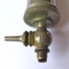
Surgery |
||
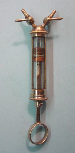
Pump, aspirating (2) by POTAIN |
||
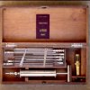
This aspiration syringe done by POTAIN comes from the estate of the physician Paul HETTO, who was established in Diekirch in 1923 - a sign of the widespread use of the syringe. He had acquired the box in Munich from the Royal Bavarian purveyor Carl Stiefenhofer. |

Surgery |
|||
Pump, aspirating (3) by DIEULAFOY |
|||
To puncture caves and abscesses served the two-way syringe after Paul-Georges DIEULAFOY (1839-1911).
Lit. G. Dieulafoy, Etude sur l'appendicite, La Presse Médicale 1896, p.12. G. Dieulafoy, Traité de l´aspiration des liquides morbides. Méthode médico-chirurgicale de diagnostic et de traitement. Paris, Masson and London, 1873. 483 pages. G. Dieulafoy, Manuel de pathologie interne. Paris, 1880-1884. G. Dieulafoy, Cliniques médicales de l'Hôtel Dieu, Paris (1897) G. Dieulafoy, Histoire de la Médecine par Maurice Bariéty et Charles Coury , Fayard Editeur. Ostini, S., L’aspirateur souscutané de Georges Dieulafoy (1869), in: Rev Med Suisse Romande. 1993 Jan;113(1):69-70. |
Surgery |
||
Pump, aspirating (1) by POTAIN |
||
Pierre Carl Edouard POTAIN (1825-1901) is better known to us as the inventor of the Sphygmo-graph named after him (see Internal Medicine). Studied in Paris. 1846 "external" from 1849-52, 1853 doctorate, 1856 senior physician in the clinic of BOUILLAUD, 1857 "agrégé", 1876 professor of internal pathology. Tragedy of Fate: POTAIN died of an aortic stenosis ...
|

Surgery |
||
A barber's razor |
||
The razor moves on the border between shaving and surgery. In the vaccination kit of Dr. med. DELVAUX has included several razors that soften the skin before it has been cut ...
1550 BC Razor arrived with movable handle, the handle was probably also used as a curler.
Quite naturally, the skin was incised by razor in the 17th century. Here is an excerpt from a report from 1665: (www.museumonline.at/1999/schools/classic/istanbul/meister.htm).
W. D. Bräutigam (published in the second edition in Weimar in 1850) wrote a "Practical Hand and Hülfsbüchlein the lower surgery for apprentices and assistants", a manual, which had been edited by the medical practitioner and surgeon Franz Wilhelm Otto Händel. Of the 231 pages of the book, no less than 31 pages deal with the use and proper handling, in particular the sharpening (removal) of razors - evidence of the importance of the razor in the daily practice of surgeons in the mid-19th century ,
The presented knife comes from the fabrication of "Coutellier" François COGNIOUL in Luxembourg (stamp on the blade). In 1849 he was naturalized (Memorial n ° 64 of 30.6.1849). In the summer of 1910, his widow took over the shop: |

Surgery |
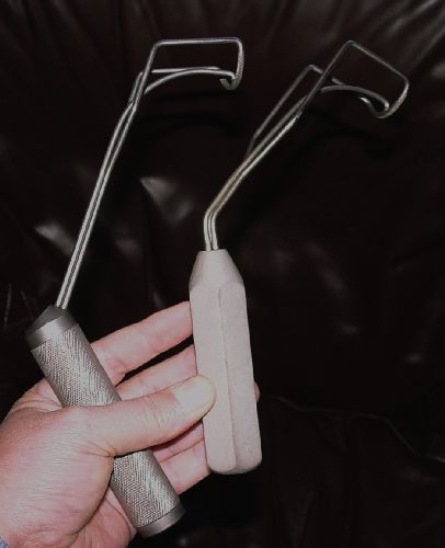
||
Retractor by COOLEY |
||
Two atrial retractors used in the Luxembourg heart surgery service of the St. Elisabeth Hospital |
Surgery |
||
Retractor by PERCY |
||
Sawing the bone at the same level as the soft tissues did not provide any material to cover the stump. CELSUS already mentions this problem: "During the operation, make sure that the remaining skin remains large enough to cover the entire stump. The Roman patient "walked on the bone", which was covered only with a thin layer of skin and at best subcutaneous fat - a painful affair if one wanted to use a prosthesis. Consequently, before cutting the bone, one tried not only to push up the skin, but also to push up all the soft tissues, thus saving a thick material for covering and cushioning the end of the bone.
Fabricius von HILDEN (1560-1634) developed a "trouser-sack", from which the stump of the bones looked down, while pulling the trouser-leg pulled the soft-tissue upwards. In the middle of the 18th century one used muscle hooks made of leather, with 2 openings for spoke and ulna resp. Tibia and fibula. These wound holders could not be cleaned. In 1800, there were the "muscle hooks" made of metal, with a single recess for the bone - illustrations can be found in Elisabeth Bennion, Old Medical Instruments, Sotheby 1979 pp. 52 and 53.
In 1799 Pierre-François PERCY (1754-1825) developed a new retractor for the soft tissue package, which consisted of two almost identical parts articulated via a hinge. He had participated in the Napoleonic campaigns where he had collaborated with LARREY - so he knew what he was talking about when he spoke on issues of amputation.
"Complete amputation for amputation", as offered by German department stores around 1910 do not contain retractors - apparently, the retractor was more a French thing. Even today, the "Percy" with removable handles part of amputation sets - new price around 157 euros!
The retractor presented here comes from the discarded fund of the clinic Differdingen (which is only a few kilometers from the French border). As in the 18th century models, there are two openings here - the small one is square, the larger one is circular and has a diameter of 4 cm. No manufacturer information, only a size "2" on the screwed-on single handle. |

Surgery |
||
Saw by HEY |
||
Trepanation saw, (according to seller 1840-90), acquired 1/2017. Of all the HEY saws I know, it is the ugliest: neither wood nor mother of pearl handle, no waist. And yet: I love my grate arbor from Romford in the county of Essex, because her last owner growled at me so irresistibly, "yes I sell abroad, but like so many, you will not buy yet".
Here is a nice paper from JAMA: "William Hey was born at Pudsey near Leeds, Yorkshire, England, the son of a respectable tradesman noted for his overbearing honesty and ramrod integrity. His maternal grandfather and great-grandfather were surgeon and physician respectively. His mother, whose good judgment usually prevailed, held the greater influence in building the character of the large family. At the age of four young Hey had an accident with a penknife and lost the sight of his right eye; however, he retained excellent acuity in the left eye, even late in life performing the details of surgical practice without the aid of a corrective lens. Before he reached the age of eight, he was enrolled in an academy near Wakefield where he was instructed in the classics, natural philosophy, and the French language. At the age of 14 he complied with the wishes of his parents and was apprenticed to surgeon-apothecary Dawson in Leeds; he remained there until 1757, when he went to London to complete his medical education. During the first winter Hey spent long hours in the dissecting room and subsequently became a pupil of Bromfield in surgery, Donald Monro in medicine, and MacKenzie in midwifery at St. George's Hospital. Having completed his formal training, Hey returned to Leeds, set up practice, and without delay acquired a reputation as a surgeon. Leeds at that time lacked facilities for hospital care which prompted him to promote a program to correct the deficiency. In 1771, he witnessed the opening of Leeds Infirmary for the admission of patients. Hey was appointed senior surgeon at the Infirmary, a position held from 1773 to 1812. In the meantime he established a close friendship with Joseph Priestly, who then lived at Leeds and who sponsored his Fellowship in the Royal Society of London which was approved in 1775. Hey was president of Leeds Literary and Philosophical Society in 1783, and was twice mayor of the city. He loved music, was a profoundly religious man, and a strong Methodist until 1781, when he joined the Church of England and wrote Tracts and Essays, Moral and Theological, including a Defence of the Doctrines of the Divinity of Christ, and of the Atonement. During his mayoralty his severe denouncement of profanity and vice led the population to burn him ineffigy. Hey suffered a series of family tragedies; three sons, two daughters, and a daughter-in-law died from pulmonary tuberculosis. Hey was an excellent surgical operator and, as a member of the Royal College of Surgeons of London, late in life gave courses in anatomy on the bodies of executed criminals at Leeds Infirmary. He introduced significant improvements in the treatment of hernia, cataract, and dislocations; suggested amputation of the foot distal to the tarso-metatarsal joint; described and named the growth, and offered evidence of the transmission of venereal disease to the fetus in utero. In addition to Hey's religious tracts he contributed several manuscripts on structural anomalies to the Philosophical Transactions and wrote a monograph, Practical Observations in Surgery, first published in 1803. His two most important contributions to clinical surgery appeared in this volume. The of the of scrotal hernia description types in infants began with deductions from the autopsy findings of an 18-month-old child. I found that the tunica vaginalis was continued up to the abdominal ring, and inclosed the hernial sac, adhering to that sac by a loose cellular substance, from the ring to within half an inch of its inferior extremity. The fibres of the cremaster muscle were evident upon the outside of the exterior sac, or tunica vaginalis. The interior or true hernial sac was a production of the peritoneum as usual, and contained only the caecum or head of the colon... Having removed the proper hernial sac, I examined the posterior part of the exterior sac, and found it connected with the spermatic vessels in the same manner as the tunica vaginalis is, when the testis has descended into the scrotum. An additional proof, that the exterior sac was the tunica vaginalis. From all these circumstances it is evident, that this hernia differed both from the common scrotal rupture, in which the hernial sac lies on the outside of the tunica vaginalis; and also from the hernia congenita, where the prolapsed part comes into contact with the testicle, having no other hernial sac besides the tunica vaginalis. To understand the cause of the hernial sac being in contact with the testicle, and surrounded by the tunica vaginalis, it is necessary to consider the manner in which this coat of the testicle is originally formed. In the foetus a process of the peritoneum is brought down, through the ring of the external oblique muscle of the abdomen, by the testicle as it descends into the scrotum; which process forms an oblong bag communicating with the cavity of the abdomen, by an aperture in its upper part. This aperture is intirely closed at, or soon after, birth. The upper part of the bag then gradually contracts itself, till the communication between that portion of it which includes the superior and greater part of the spermatic chord, and the lower part of the bag, which includes the testicle and a small share of the chord, is obliterated. The lower part of the process or bag retains its membranous appearance, and is called tunica vaginalis testis propria; while the upper becomes an irregular cellular substance, without any sensible cavity, diffusedamongst the spermatic vessels, and connecting them together. In the hernia which I am describing, the intestine was protruded after the aperture in the abdomen was closed; and therefore the peritoneum was carried down along with the intestine, and formed the hernial sac. It is evident also, that the hernia must have been produced while the original tunica vaginalis remained in the form of a bag as high as the abdominal ring; on which account that tunic would receive the hernial sac with its included intestine, and permit the sac to come into contact with the testicle. The proper hernial sac, remaining constantly in its prolapsed state, contracted an adhesion to the original process of the peritoneum which surrounded it, except at its inferior extremity: there the external surface of the hernial sac was smooth and shining, as the interior surface of the tunica vaginalis is in its natural state. This kind of scrotal hernia may, therefore, not improperly be called hernia infantilis, as it can only exist when the rupture is formed while the parts retain the state peculiar to early infancy. The scrotal hernia may be divided into three species, the specific difference of which arises from the state of the tunica vaginalis at the time of the descent. If the abdominal aperture of this process is open when the intestine or omentum is protruded, the rupture is then called hernia congenita. If the upper part of the process remains open, but the abdominal aperture is closed, and is capable of resisting the force of the protruding part, the hernia then becomes of that species which I have now described, the hernia infantilis. If the cavity of the upper part of the process is obliterated, and the septum is formed a little above the testicle, as in the adult state; the hernial sac then descends on the outside of the tunica vaginalis, and forms the most common species of scrotal rupture, which may with propriety be called hernia virilis. In the same treatise Hey discussed the factors involved in internal derangement of the knee that follows minor trauma. The disease is, indeed, now and then removed, as suddenly as it is produced, by the natural motions of the joint, without surgical assistance: but it may remain for weeks or months, and will then become a serious misfortune, as it causes a considerable degree of lameness. I am not acquainted with any author who has described either the disease or the remedy; I shall, therefore, give such a description as my own experience has furnished me. The leg is readily bent or extended by the hands of the surgeon, and without pain to the patient; at most, the degree of uneasiness caused by this flexion and extension is trifling. But the patient himself cannot freely bend, nor perfectly extend the limb in walking; but is compelled to walk with an invariable and small degree of flexion. Though the patient is obliged to keep the leg thus stiff in walking; yet in sitting down the affected joint will move like the other. The complaint which I have described may be brought on, I apprehend, by any such alteration in the state of the joint, as will prevent the condyles of the os femoris from moving truly in the hollow formed by the semilunar cartilages and articular depressions of the tibia. An unequal tension of the lateral, or cross ligaments of the joint, or some slight derangement of the semilunar cartilages, may probably be sufficient to bring on the complaint. When the disorder is the effect of contusion, it is most likely that the lateral ligament on one side of the joint may be rendered somewhat more rigid than usual, and hereby prevent that equable motion of the condyles of the os femoris, which is necessary for walking with firmness".
|

Surgery |
||
Scalpel, surgical (1) |
||
This scalpel with the obstruse handle "Manual Tool Cie Sheffield" I bought 1998 at a flea market in Canada - maybe a visitor of these pages can tell me something about the form and function of the handle ...
An identical object was offered in 2005 by "jayart3" as a scalpel of the company WRAGGS on the English EBay market: "A 19th Century surgeon's scapel by Wraggs of Harwood Street, Sheffield." The maker mark is shown on the handle rosewood with a Sheffield steel blade and a black cover The blade is 6cm long and the black cover is 8.5cm long ".
A company WRAGGS is still in Sheffield today, but "electric".
Nota: to the so-called eraser knives: Langenbeck speaks literally of the "eraser of the convex scalpel" (Conrad Johann Martin Langenbeck, Nosology and Therapy of Surgical Diseases, Göttingen 1830 Volume 4, 4th section p.163). |
