-
Home

-
Human Medicine
-
Surgery
- Artikel


")
Chirurgie |
||
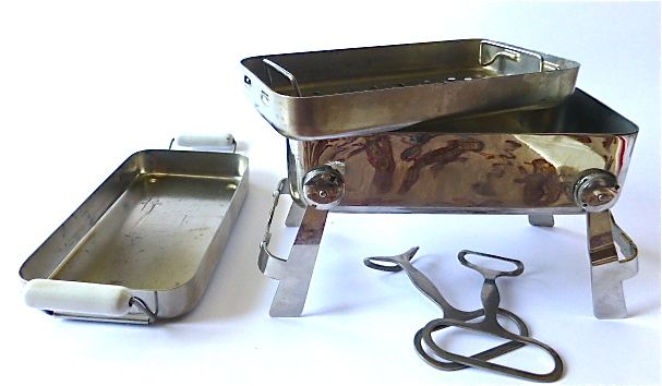
Sterilizer (4) |
||
We present a small (17 x 8.5 x 4.5 cm when folded) pocket sterilizer, a gift from Dr. Fritz Teichner from Wetzlar. A big thank you from this point.
You can see the lid (left in the picture) with the two handles made of porcelain - just like the big models. Two hooks (in the picture below the stove) are used to remove the bowl (in the picture on the top of the cooker) on which the item to be sterilized lay. Two retractable feet. No manufacturer information. The spirit stove is missing, but has left behind on the underside of the stove clear heat traces.
This type of sterilizer goes back to the surgeon Curt SCHIMMELBUSCH, which is why I introduced the device in the chapter on surgery, even though these small specimens have been used many times by internists to sterilize their syringes and implants. |
Surgery |
||
Sterilizer (5) |
||
From the estate of the doctor Guillaume KOENER, who was established in Clerf in 1913, comes the aluminum jug presented here, the bottom of which is removable, with three openings concealed by a linen cloth.
The 16 cm high pot, with a diameter of 8 cm, was used to sterilize small objects in hot water. |

Chirurgie |
||
Sterilizer (3) |
||
From the estate of Dr. Guillaume KOENER / Clerf. |

Surgery |
||
Surgical kit (1) |
||
Tweezers In the middle of the sixteenth century, the tweezers appeared on the instrument table of the Florentine Vidus Vidius (1509-1569), also known as Guido Guidi, drawn by Francesco Salviati (Chirurgia e Greco in Latinum conversa, 1544).
A Henri Mongin (* after 1766 in Crepey, died before 1843), son of a Henri Mongin and Cathérine Perdouche, married in 1791 in Saint Benoît a M.-Antoinette Naze. Whether he is the wanted company founder, I can not say. In any case, the company Mongin was founded in 1814 by a certain Nicolas Mongin and since then has been making knives - it is now part of a larger group called "Groupe Mongin", in which the companies FAURE, GRINCOURT, LUM and SAOTM are combined.
|
Surgery |
||
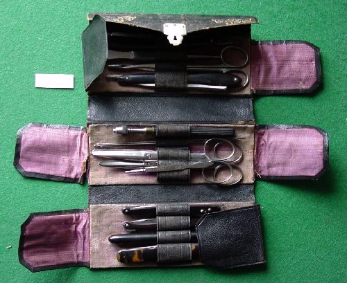
Surgical kit (2) |
||
Empty folding bag (French "trousse de chirurgien", "folding pouch"), bought at a flea market in Arlon (Hall polyvalent) on 27.1.2007. On the inside of the hinged lid the stamp of the company F. SCHWOB, successor of the company VITRY frères.
|

Surgery |
||
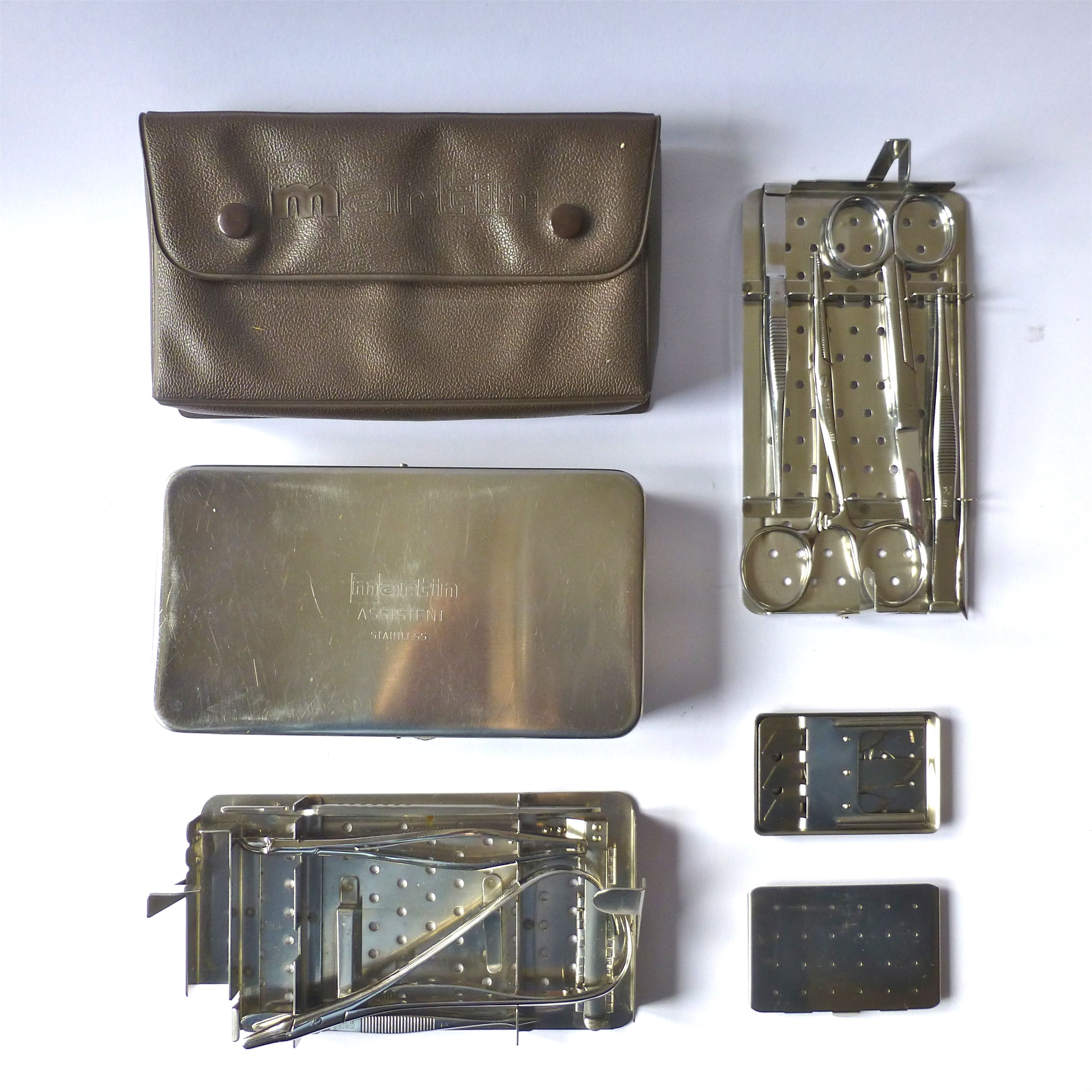
Surgical kit (3) |
||
From the suitcase of Paul ROLLMANN, a physician established in Bonneweg / Luxembourg, came this small suture set: a metal container with its plastic case from MARTIN (model "assistant standard", catalog 1982 p. 295) with two inserts on which cutting and suture devices (scalpel , Tweezers, scissors, probe, clamps, round needles and needle holders) were stored in pre-punched incisions.
|
Surgery |
||
Syringe Rotanda by JUENGLING |
||
In 1920, the company Wilhelm Haselmeier GmbH & Co got its start in a small backyard workshop in Stuttgart-Degerloch. In 1925, the founder Wilhelm Haselmeier developed the Rotanda reusable syringe, which is still produced almost unchanged to this day.
Originally intended as a "three-way" syringe for blood transfusion, there was quickly a "two-way version", with the liquids out of the body - but could also be pumped into the body - a "German syringe to Dieulafoy" so to speak (see Chapter Surgery under aspiration syringes).
Presented is such a two-way version.
Otto A. JUENGLING (1884-1944) had studied in Tübingen and Kiel and had been an assistant at the Pathology Institute under Prof. Paul Clemens von BAUMGARTEN (1848-1928), later at the Surgical Clinic of Prof. PERTHES in Tübingen. Habilitation 1919, associate professor 1923. From 1926 head of the surgical department at the Municipal Katharinen Hospital in Stuttgart. "When the Tübingen surgeon Otto JÜNGLING got to know the syringe design of the Stuttgart instrument maker Wilhelm Haselmeier in the 1920s, he was immediately enthusiastic: the often-used, cumbersome two-way or three-way stopcocks are now expendable, and their function is now taken over by the specially designed" head piece ". Depending on the position of the rotatable syringe body, it releases one of several ways, the selected setting marks an arrow on the glass cylinder.
|

Chirurgie |
||
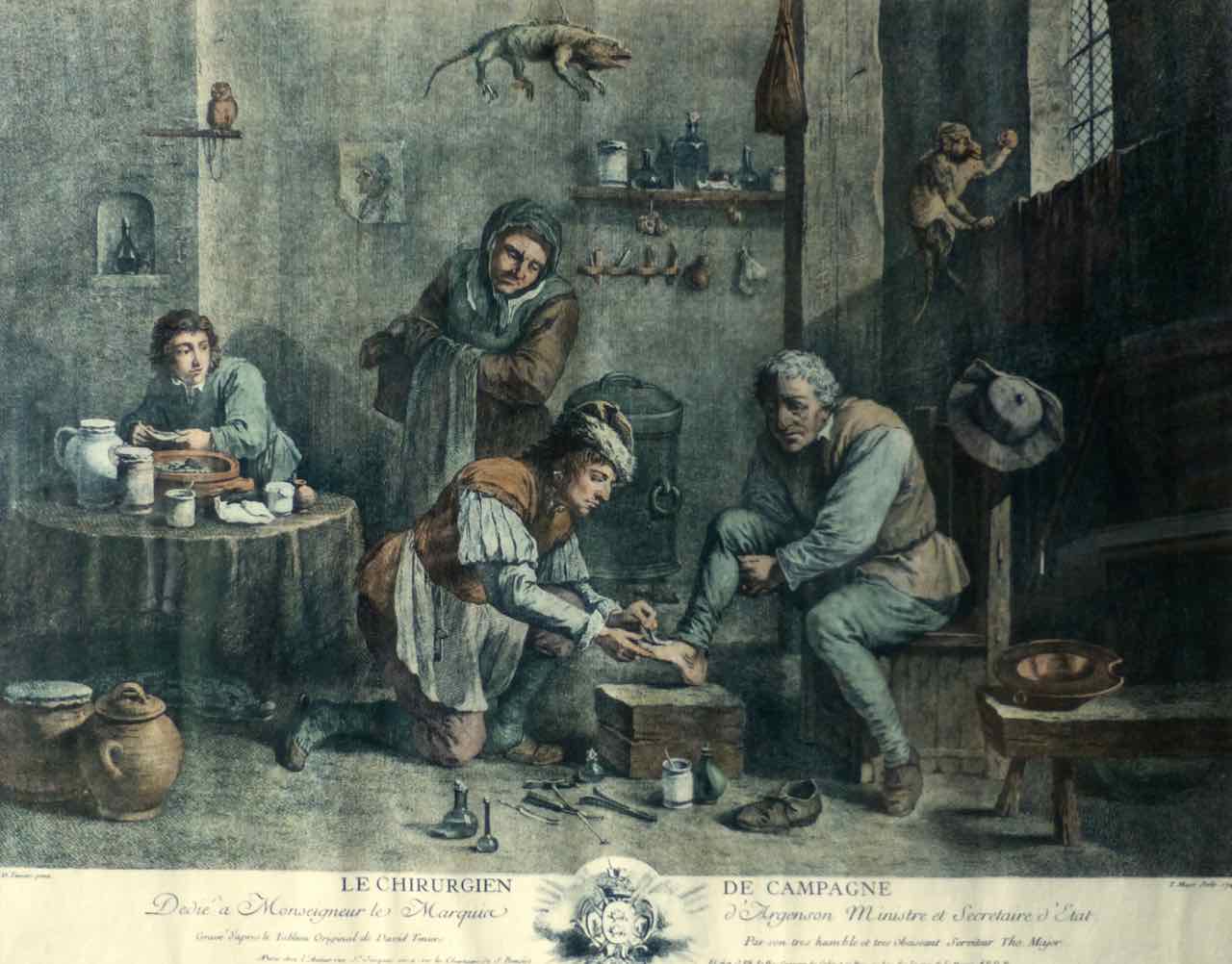
Teniers, The village surgeon |
||
"The country surgeon". Colored print of an engraving, done in 1747 after a painting by the Flemish painter David TENIERS the Younger (1610-1690) by Thomas Major (1720-1799). Treatment room of a surgeon, animals stuffed on the walls and alive, ointment pots and instruments, on a stool a man whose foot is treated. In the foreground surgical instruments of that time: Pelikan, Zahnzange, lever ...
One can argue whether more surgeons or more pedicurists - the last term earliest occupied for the year 1762 "Rousselot, chirurgien de Mgr le Dauphin, the Princes et de Mesdames, en cette partie, ancien chirurgien de M. le prince de Wirtemberg, toilet of the pied, ou traité de la guérison des cors, verrues et autres maladies de la peau, 1762 ".
About David TENIERS TENIERS received his master's degree in 1633 and was admitted to the Luke Guild of the City of Antwerp, where painters as well as surgeons were incorporated - hence free access to the workplaces of his colleagues. 1645/46 he held the post of Dean of the Luke Guild. Among other things, TENIERS created peasant picnic scenes with drinkers, smokers and card players, as well as motifs such as folk festivals, soldiers' parlors, surgeons, bathers, alchemists' and witches' kitchens - a total of 800 works have been preserved. Among his patrons we find the Austrian governor (1746-1756) in Brussels, Archduke Leopold Wilhelm (1614-1662), youngest son of Emperor Ferdinand II and founder of the collection of paintings of the Kunsthistorisches Museum in Vienna. TENIERS portrayed him in 1651 in the middle of his collection of paintings in the Palais de Bruxelles (now Kunsthistorisches Museum, Vienna). For the medical historian, the interest of the Archduke (as well as his brother, Emperor Friedrich III) to alchemy or gold making is worth mentioning. In 1656 he set up a laboratory "above the Ballhaus" in Vienna.
47x32.5 cm large picture, gift of a patient. |
Surgery |
||
Tenotomy scalpel |
||
"Ténotomy, s.f. Syn. Ténontotomie, opération qui consiste à sectionner un tendon pour redresser un membre, un segment de membre (pied bot) ou un organe (œil atteint de strabisme). On applique également ce terme à la brides section fibreuses cicatricielles qui gènent certains mouvements ou maintiennent un membre ou un segment de membre en mauvaise position »(M. Garnier, Dictionnaire des termes techniques de médecine, Paris 1912).
The experience during the Napoleonic campaigns had the French surgeons make bold. The research was fostered by the problem of "clubfoot" by several prominent patients
For the intervention of the tendon separation writes the Larousse médical 1924: Excessive tenotomies eventually led to the discredit of the tenotomes ... |

Surgery |
||
Thoracal drain by BÜLAU |
||
Already the Parisian internist Pierre-Charles Ed. POTAIN (1825-1901) had devised an aspiration apparatus. The lung specialist (surgeon and internist) Gotthard BÜLAU (1835-1900) was supposed to improve him.
biography Gotthard BÜLAU was born on February 27, 1835 in Hamburg, the son of the physician Gustav Bülau (1799-1857). From 1854 studies in Heidelberg and Göttingen, where he received his doctorate in 1858. In the fall of 1858 he became a medical assistant at the General Hospital St. Georg, and stayed here for three full years. In the spring of 1867 he became a representative for the diseased chief physician of the house, Dr. med. Georg Karl Franz TÜNGEL (1816-1873). As a volunteer doctor remained BÜLAU in the hospital until 1869, and then, after retiring TÜNGEL's, chief physician of one of the four sections of internal medicine of the house. He remained chief physician until 1886, then gave up the hospital post, as his increasing private practice made him impossible this activity. In 1888 he practiced "Alsterterrasse, 10, 1897 Mittelweg 13". BÜLAU died on October 20, 1900.
principle At St. Georg's Hospital in Hamburg, he devised a "water lock", a continuous suction device for emptying pleural cavities, which works on the lifter principle. In 1875 he combined a chest drainage with permanent suction and healed a pleural empyema. His apparatus is "neither a special tube or catheter, nor a bottle or a localization, but the principle of perpetual suction!" (Dr. med. Thomas Kiefer).
Indicated is the preparation of the device in patients, in which (after surgery and after inflammation) the pleural space with air or liquid (blood, serum, pus) has filled (pleural empyema). With the device you could aspirate liquid, but also pump gas into the chest. This therapy, known as "collapse therapy", was the best treatment option for pulmonary tuberculosis around 1900. The artificial pneumothorax was described in 1882 by the Turin practitioner Carlo FORLANINI (1847-1918). The Marburg surgeon Ludolph Brauer (1865-1951) introduced the method in 1906 in Germany. While spontaneous pneumothorax is not uncommonly fatal, astonishing success has often been achieved with this controlled collapse of the lungs. "Through a system of two communicating tubes filled with water, underpressure or overpressure can be generated, so that air can be filled or sucked into the pleural space by means of a puncture needle connected to a tube, resulting in a healing of the affected patients Lung.
The procedure has long played a prominent role in the treatment of pulmonary tuberculosis and was replaced only after the Second World War by the introduction of antibiotic therapies "(Michel Martin, Institute of Medical Ethics and History of Medicine at the Ruhr University Bochum).
technology Introduction of a drainage needle via a guide spike in the 2nd or 3rd ICR parasternally.
exhibit A wooden box measuring 58x40x16.5 cm will be presented, which was used until the 1970s in the St. Elisabeth Clinic in Luxembourg. Manufacturer: Fa. P.A. Stoss' successor in Wiesbaden, Inh. Max Helfferich - a specialized company founded in 1892 that still exists today at the Kreuzberg Ring 36 in Wiesbaden. Link:
Lit.: - Paul E. Van Schil et al., Thoracic Drainage and the contribution of G. Bülau, in: Ann. Thoracic Surg. 1997, 64:1876. |

Surgery |
|||
Thread box |
|||
The box presented here with telescopically extendable compartments contained a collection of threads in the original case when purchased (Metz-Grigy, April 16, 2005) in the upper drawer. All other shops were filled up with "Wattepäckchen".
Because of the threads we want to introduce him to the collection at this point. From top to bottom can be found in the upper drawer: - Drain stérilisé n°16 Laboratoires Larochette - Villefranche s / S Paris. - Crino Larochette n°2. - Crins de Florence stéril. moyens - Catgut Stéril. n°2 Clas.dec. n°6. Diametres entre 0mm60 et 0mm69 - Soie stérilisée n°2 - Crins moyens - Catgut stéril. n°1 Clas.dec. n°5. Diametres entre 0mm50 et 0mm59
Exhibit Plastic tipping bottle, Perlon thread braided O |

Surgery |
||
Thread, so called caterpillar |
||
From the FANDRE works in Nancy comes the curious, now abandoned material "silkworm good" (Crins de Florence).
Gustav KLEIN reported in 1907 on his very positive experiences with the silk seam in laparotomies (Zbl.Gyn.Nr.33 1907 S. 1004). R. Benndorf also praised the advantages of the material (Zbl.Gyn. 1907 No. 51 p. |
