-
Home

-
Human Medicine
-
Surgery
- Artikel


")
Surgery |
||
Thread from horse hair |
||
Horse hair is one of the earliest suture materials - tear-resistant, available in considerable length and in large quantities, it was already used by the ancient Chinese to suture wounds, so In a Chinese script from the 3rd century BC Chr. Is reported by a doctor named HUATO, who was able to remove diseased organs and thus heal people. An extraordinary note, since the then Chinese medical tradition normally prohibited surgery. Perhaps, according to the researchers, this huato legend refers to a medical tradition of mummies unearthed in Xinjiang province, as these people have undergone surgery, as evidenced by the discovery of a surgical incision sutured with horsehair.
The ancient Egyptians used ant pliers to adapt to skin wounds - a method they may have adopted from the Indians. The Greeks preferred fine tendons as sutures.
The Arabs introduced the suture with strings of musical instruments for deep seams - and horsehairs for skin sutures. Later, various materials were used: dried animal casings and tendons, skin cut into strips, maiden hair, birch bark strips, hemp and grasses. In the Middle Ages, one took horse or human hair and made a strand of it; This was inserted into an incision of the skin (calf, neck or groin) so that both ends of the strand protruded; these were knotted so that the strand did not slip out. The purpose of this case was to stimulate the antibodies (because the incision naturally inflamed) and thus more defenses against diseases (such as the plague) were available ...
During the American Civil War in 1865, different sutures were used: - Surgeons of the Northern States used (unsterile) "silk" silk, - southerners used cotton, - Since catgut was excessively expensive, the Confederates often used horsehair at the end of the war. Since this was annoyingly rigid, you cooked your hair to soften it. The suture material was sterilized without anyone having been aware of this fact. The wounds healed cleaner and faster than before ...
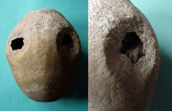
Gustav SIMON (1824-1876), professor of surgery in Rostock and Heidelberg, introduced the seam with horsehair into the operating theaters of Europe ... Still today, mane hair is used by horses in African folk medicine, such as "female genital mutilation" - as a suture in this despicable intervention sheep, horse hair, acacia thorns, raffia or iron rings and for hemostasis ashes, herbs, cold water, leaves and wound compresses Sugarcane used. The "horse-hair" bottle presented here contains 2 strands of 20 inches (x2.54 = 50 cm) each. It comes from a US plant of the pharmaceutical company JOHNSON & JOHNSON and was increased in January 2005 on Ebay. As a specialist in surgical sutures, the success story of the JOHNSON & JOHNSON subsidiary Ethicon began 40 years ago. Headquarters is Hamburg Norderstedt. There are currently 1,500 employees here. |

Surgery |
|||
Traction bows by KIRSCHNER |
|||
Lit.: Kirschner, Martin & Stauss, Beiträge zur willkürlichen Begrenzungn und zur individuellen Dosierung der Spinalanästhesie. Erfahrungen an über 1000 Fällen (S.322-383, 26 Abb.), in: Verh. Dtsch. Ges. Chir., 56. - Berlin, Julius Springer, 1932, 8°, CXVIII, 856 S., 260 Abb. Kirschner, Martin: Die Hochdrucklokalanästhesie. Berlin, Springer-Verlag, 1944, 8°, 62 S., 32 z.T. farb. Abb., OKartBd. Erste Auflage der letzten und posthum erschienen Arbeit von Martin Kirschner hrsg. v. R.Zenker Weißer, Christoph: Die Knochenbruchbehandlung bei Martin Kirschner und die Entwicklung des „Kirschnerdrahtes“. Anmerkungen zu einer genialen Idee in der Chirurgie, in: Würzburger Medizinhistorische Mitteilungen 12 (1994) S. 5-18. |

Surgery |
||
Thread silk (3) |
||
Dr. VÖMEL's original sewing material
The 6.5 resp. 7.2 cm high vials contained silk of varying caliper (N ° 3 / N ° 4).
Towards the end of the 19th century the "German School" left the use of silk. August MARTIN writes:
To the manufacturer In 1891 Gottfried VÖMEL founded a surgical suture material factory in Kronberg / Taunus - the company still exists today and mainly produces surgical sutures and dental floss. |

Surgery |
||
Trepan, cranial by DOYEN |
||
As a special master of trepanation, the surgeon Roger FRUGARDI from Salerno proved in the second half of the 12th century. He had developed a method of sawing his patients out of the skull with great precision. His work "Practica chirurgiae", published around 1180, set new standards for the guild.
Until the invention of the spherical drill by DOYEN in 1895 the Krontrepan remained in use. "Trepan à cliquet" resp. "vilebrequin" with ball-mill after Eugène-Louis DOYEN (1859-1916).
About the person of DOYEN
Indications of trepanation "Hollerich, June 22. Last night, the worker Debras from Hollerich was hit in the local steelworks by a falling slag on the head, and the unfortunate was transported to the clinic at the fish market, where this morning a cranial operation was carried out, which went well However, not all Gefabr has been eliminated yet: Debras is married and the father of a child "(LW of 22.6.1906). A forgotten indication is aneclamic seizures during pregnancy: According to Wilhelm ZANGEMEISTER (1871-1930), the trepanation in eclampsia can be life-saving!
Exhibit Crank drill by Fred HASLAM in New York, Boring mill by Vincenz MULLER in Chicago Origin: Ebay, import from the USA
Link
|

Surgery |
||
Trepanned cranium, Peru |
||
The trepanation - the opening of the bony skull - was the first major surgical procedure people performed. Since the days of the country doctor Pierre Barthélémy PRUNIERES (1828-1893), who discovered skulls in prehistoric skulls in the Lozère valley in southern France in 1873, one knows that skulls were traversed in ancient times. His compatriot Paul BROCA (1824-1880) recognized that these had been done on living and surviving patients. In 1874, at an anthropological congress in Lille, Pruneier showed him one of his skulls, and Broca immediately recognized the callus at the margin of the trephine opening, a testimony to the survival of the operated person.
The survival rate of the trephines varied in the Neolithic period depending on the surgeon:
Three problems had to be solved for the surgeon:
Magic rites or rational treatment of skull injuries? After initially thinking of magical backgrounds, especially in prehistoric trephines, recent studies on craniofacial surgery in Peru show residual states after trauma in the majority of skulls. Apparently, the trepanation corresponds to a special kind of warfare of that time, when you slashed your opponent with a sling and stone-honed club ...
exhibit The presented skull comes from the disbanded collection of a US diplomat who acquired the skull in 1950 by an anthropologist in Lima. Presumably the peninsula PARACAS, where the researcher Julio C. TELLO (1880-1947) from 1925 excavated hundreds of these skulls. The so-called Paracas civilization existed from 800 BC. until about 200 AD. One reason for their disappearance, the archaeologists could not make out to this day. Similar skulls were recently found among the people of Chachapoya, the "cloud people", who lived in the period 800-1475 AD. lived in the high mountains of northern Peru. From the Paracas they had apparently learned the technique of trepanation in scraping technique.
The earliest skull opening technique is probably the so-called "area cockroach". In Peru, it was scraped but also sawn, chiselled and drilled. The scraper technique was applied to the skull presented here with its typical double reworking. After the recordings, we wiped away the cracks on the surface of the skull - they testified to the particularly brash manner of some of our contemporaries to deal with the remains of their ancestors. But we did not want to issue these "autographs" in our small museum, but the skull with its finely healed wound edges.
Lit .:
If you want to treat yourself to the "kick" of a trepanierten skull on holiday, go to Normandy to Avranches. Here, in the basilica "Saint-Gervais-et-Saint-Protais", the skull of Sts. Aubert was exhibited as "Le crâne avec l'emprinte du doigt de l'archange Michel" - in reality a Stone Age skull with traces of a trepanation. Recent investigations have also raised the hypothesis of a skull cyst in a medieval skull ...
Our ancestors, the Celts, first practiced the scraping technique. Like a noble luxury item, they eventually imported a new trepanation method from the Mediterranean to the north: drilling with a hollow drill, which cut a round bone plate from the skull. Not infrequently, these skull slices were worn in ancient times as an amulet around the neck |
Surgery |
||
Trephine, cranial |
||
"Lamadeleine. 3. Okt. Der Feldhüter Libert Joh. erhielt am verflossenen 19. Aug., Abends auf offener Straße, von einer unbekannten Hand, den Schädel, eine halbe Handbreit, eingeschlagen. Trotz seiner schrecklichen Wunde wurde dieser Unglückliche während sechs Wochen am Leben erhalten, und bereits stand er von seinem Bette auf, um mit den Seinigen an demselben Tische zu essen, als sein Zustand sich in den letzten Tagen so sehr verschlimmerte, daß an sein Aufkommen nun mehr kaum zu denken ist. Die zwei Aerzte Hr. Dr. Lehnertz aus Rodingen und Hr. Dr. Stronck aus Aubange, haben, diesen Morgen die äußerst seltene und gefährliche Operation der Trepanation als letztes Rettungsmittel an ihm vollzogen" (L.W. vom 5.10.1878).
In 1885 a joung boy was trephined in Luxembourg
He recovered and died in 1893 (L.W., .5.1893).
"Das Instrument, mit dem man ein rundes Stück aus dem Knochen ausbohrt, nennt man Trepan (Trephine); sein gezahntes, einer Kreissäge von etwa 1½ cm Durchmesser entsprechendes Ende heißt die Trepankrone. Das ausgesägte Knochenstück wird mit einem hebelartigen Instrument (Tirefond) herausgehoben und sodann der Fall je nach seiner individuellen Beschaffenheit weiter behandelt. Schon im Altertum, namentlich in der Kriegschirurgie, sehr häufig vorgenommen, gehört die T. jetzt zu den selten zur Ausführung kommenden Operationen, da sie früher außer bei Verletzungen auch bei Geisteskranken ausgeführt wurde (Wilhelm v. Saliceto). Auch das Brustbein hat man trepaniert, namentlich um Eitermassen, welche sich hinter demselben entwickelt hatten, zu entfernen. Unter allen Umständen ist die T. eine lebensgefährliche Operation, weil sie zu einer schweren ältern Verletzung eine nicht minder schwere neue hinzufügt" (Meyers Konversationslexikon).
Exhibit Monnier sold the "Tréphine à Manche avec sa couronne". The same model was also distributed by the company Esculape. |

Surgery |
|||
Trocar (1) |
|||
A trocar (Latin for "acus triquetra", triangular needle) is a dagger-like needle with handle and triangular point.
Trocar with central enema - above: ready for puncture - below: with withdrawn engraver
The trocar presented here with wooden handle and "spit-shaped" spout originates from the "Metzer Wunderkiste" and was probably in use at the end of the 19th century |

Chirurgie |
||
Trocar (2) |
||
Santorio SANTORO (1561-1636), who taught in Padua and later in Venice, and gained significant knowledge in the field of physiology, invented the trocar for paracenthesis of the abdomen.
This invention was particularly important at a time when there were no effective diuretics to treat the ascites.
Johann Collins WARREN 1778-1856), professor of surgery in Boston / USA, was the first to have the courage to puncture the pericardium. |

Surgery |
||
Trocar (3) |
||
Stainless steel trocar with side inlet.
Even today, the trocars are used to evacuate massive effusions not only in the abdomen but also in the pleural space and in joints. |

Surgery |
||
Trocar (4) |
||
|

Surgery |
|||
Trocar (5) by VERESS |
|||
The VERESS needle (also known as the VERRES needle) is a special kind of trocar: every laparoscopy begins with the insertion of the VErRES needle.
The first laparoscopy in humans was performed in 1910 by the Swede Hans Christian JACOBAEUS in Stockholm. In 1933 VEWERS first filled carbon dioxide into the abdominal cavity instead of air. This has the advantage that residual gas in the stomach are painlessly absorbed by the body and that you can easily work with electrical instruments.
The assistant of the Surgical University Hospital Halle Otto GÖTZ (X-ray diagnostics in gas-filled abdominal cavity, a new method) developed in 1918 an automatic special needle for the safe introduction of gas into the abdominal cavity - purpose of his method was the production of double contrast X-ray photographs.
A significant advancement of endoscopic technique was achieved in 1938, when the Hungarian surgeon Janos VERESS "rediscovered" the needle of GÖTZE and used it for a safe puncture of ascites and a safe system of pneumothorax (treatment of pulmonary tuberculosis) - he applied this needle to the 2,000 times, but NEVER used them to create a pneumoperitoneum. NEVER he suggested using them to create a pneumoperitoneum! Nevertheless, apart from minor modifications, the needle is nowadays used for the installation of such pneumoperitones!
The VERESS needle has a centrally-rounded pin rounded off at the tip, which is pushed forward by a spring as soon as the peritoneum is pierced, thus avoiding injury to internal organs. The gas is introduced via a lateral opening on the advanced central pin. Once the tip of the needle has punctured the peritoneum, the blunt stylet snaps in and prevents bowel injury. Gas is then pumped into the abdominal cavity through the laterally perforated stylet (pneumoperitonaeum): the brook covers are lifted off the intestinal loops; The surgeon can push the laparoscope into the space created in this way. One-third to one-half of complications in laparoscopy occur during insertion of the needle or the first trocar (Leonard 2000, Chapron 1998). A vascular injury caused by a Verresnadel or the first trocar is a typical, overall rare, but well-known complication of a laparoscopy, which can not be avoided even with all due care, because these instruments must be introduced without visual inspection. The injury of a vessel therefore does not indicate a faulty procedure of the physician.
As a site for the insertion of the VERRES needle, a wide variety of sites were recommended - left subcostal - the posterior vaginal vault - the uterine fundus by guiding the needle vaginally through the cervical canal (in particular, this puncture is suitable for obese women) - The lower circumference of the navel - the common puncture site. As a place of puncture, the umbilical pit is usually taken, because here the distance between the skin and the abdominal cavity is thinnest. In addition, the wound in the navel and re-stitched wound visually not later ...
Note: the Verres needle may also be advanced into the pleural space at the top of the third rib for thoracoscopy. |

Surgery |
||
Tweezer, hemostatic by BERGMANN |
||
Already CELSUS knew the vascular ligation around 35 AD - when his work was forgotten, the technique of ligature was lost. Only Ambroise PARE (1510-1590) took up the method of artery ligation in 1552 again.
- "Pinces d'AMUSSAT munies d'un cursur pour les tenir fermées" in the "Aide-Mémoire de l'opérateur" of ISNARD 1849 p. 6). Jean-Zuléma AMUSSAT (1796-1856) described in 1829 the torsion of the vessels as a suitable haemostatic method (Archives générales de médecine, Paris, 1829, 20: 606-610). Quickly took over in Germany, the technology: - the tweezers used by Johann Carl Georg FRICKE (1790-1841), the father of clinical surgery in Germany, did not yet have teeth. It served the torsion of the vessel, not the ligature:
Who had finally invented the tweezers, the German FRICKE or the Frenchman AMUSSAT, everyone wants to have been! The tweezers were later modified again and again (negligible).
- Ernst v. BERGMANN (1836-1907) added teeth to better grasp the vessels. Around 1880, b. BERGMANN plans to sterilize the surgical instruments ... In 1886 he left the chemical antisepsis, together with his Berlin student Curt Schimmelbusch, to apply steam-sterilized dressing materials.
Presented is a serrated arterial tethering forceps (engl., Artery-forceps), purchased at a street market in Arlon June 2007. N ° 32 |
